
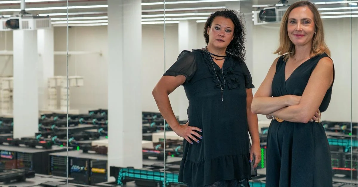
Researchers in Barcelona have built an artificial intelligence tool that tells young blood stem cells from aged ones by looking at 3D microscope images of their nuclei, according to the Bellvitge Biomedical Research Institute (IDIBELL), which led the work, and the newspaper Ara. The tool, called ChromAgeNet, is described in the journal Aging Cell, and the results were announced at the end of September. The study was done in mice.
The work was led by Maria Carolina Florian, an ICREA research professor in IDIBELL's regenerative medicine programme, and Paula Petrone, of the Barcelona Supercomputing Center (BSC-CNS) and the Barcelona Institute for Global Health (ISGlobal). It was a central part of the doctoral thesis of Pablo Iañez, an ISGlobal researcher.
Reading age in how DNA is packed
Ageing changes the way a cell organises its genetic material inside the nucleus, in a structure called chromatin. The team stained hematopoietic stem cells, the cells that produce blood and immune cells, with DAPI, a cheap and widely used DNA stain, took high-resolution 3D images and trained a convolutional neural network on them. Based on the appearance of the nucleus, the model had a 77% probability of sorting cells correctly into young and aged, better than a machine learning model built on chromatin features the researchers had defined by hand. Aged cells showed a "more disorganized" structure, in Florian's words.
The differences are not necessarily visible to the eye. Asked which features drove its predictions, the model pointed to chromatin entropy, heterochromatin located at the periphery of the nucleus and certain chromatin condensates.
A screening tool, not a cure
As a proof of concept, the team treated aged cells with epigenetic drugs and used ChromAgeNet to check whether their chromatin looked younger; with some compounds it did. The researchers stress that this does not show the cells were functionally rejuvenated. Because the stain is low-cost and the model is small, they see it fitting into high-throughput microscopy to test large numbers of compounds. They have also released a dataset of three-dimensional images of these cells along with the tool.
"The tool we have developed is important because it helps us understand the mechanisms that can be a therapeutic target," Florian told Ara (translated). The next step is to check whether the same signature appears in human cells. The work was funded by an ERC Consolidator grant, the Spanish Ministry of Science and the La Caixa Foundation.